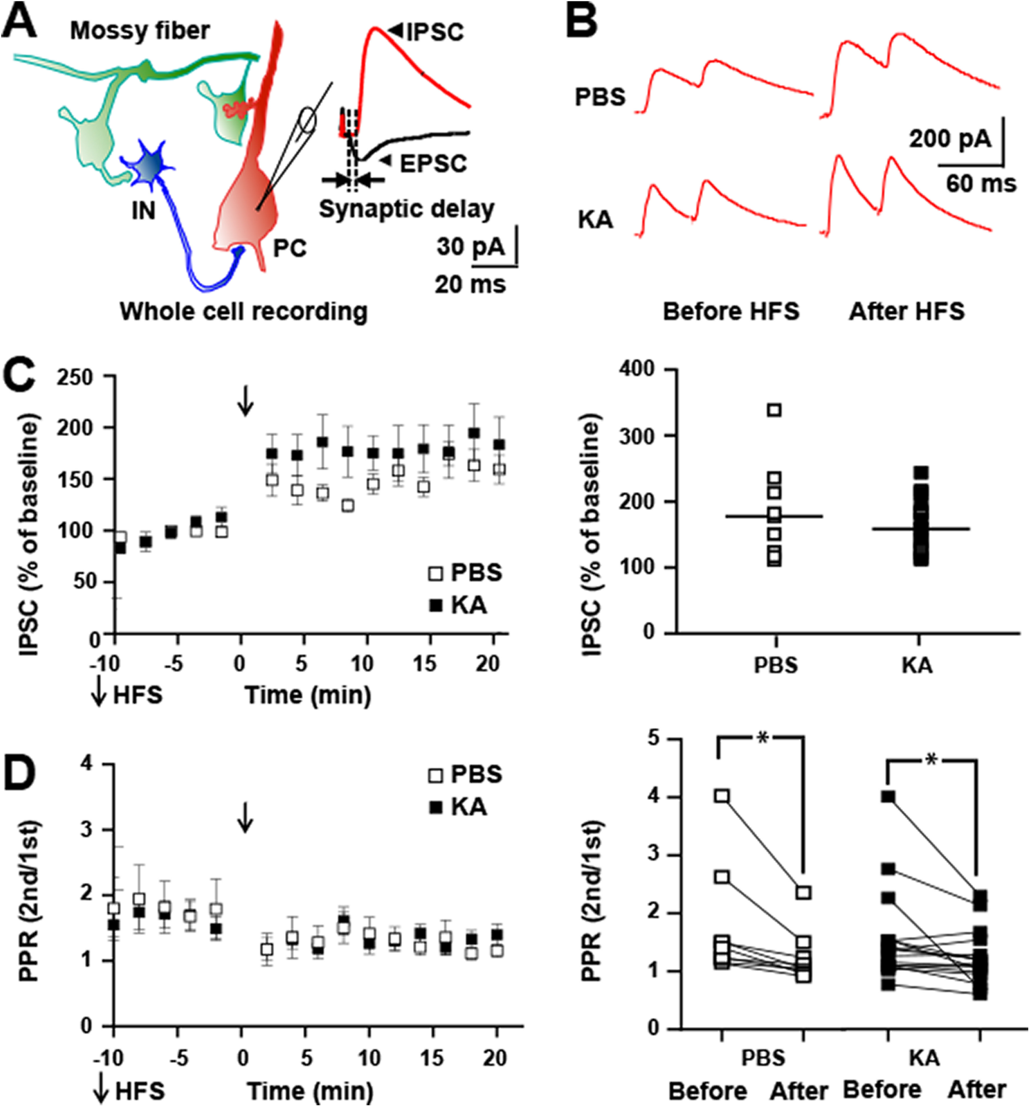
Figure 5.

HFS of mossy fibers in vitro induces LTP of feedforward IPSC in both controls and following status epilepticus. A, Schematic of local circuit (left) in which activation of granule cell evokes monosynaptic EPSC and disynaptic IPSC recorded in CA3 pyramidal cell (right). Note the delay between onset of EPSC and IPSC approximates 2.5 ms similar to Torborg et al. (2010). B, top, Representative traces show individual IPSCs recorded at holding potential of 0 mv collected 10 min before and between 10 and 20 min after application of HFS in slices isolated from PBS or KA infused mice. C, left, HFS (denoted by arrow) produced LTP of mossy fiber-CA3 disynaptic IPSC in slices from both PBS (184 ± 24%, n = 9, p = 0.0001, post hoc Bonferroni’s) and KA (164 ± 9%, n = 17, p = 0.0001, post hoc Bonferroni’s) infused animals. Right, Results of individual cells are plotted. D, PPR of mossy fiber evoked IPSC of experiment of C above. Repeated measures ANOVA revealed a p value of 0.0001. Post hoc Bonferroni’s revealed no significant differences between PBS and KA either before or after HFS. Post hoc Bonferroni’s revealed a significant reduction of PPR following HFS in both the PBS (1.76 ± 0.3 before, 1.24 ± 0.1 after, n = 9, p = 0.02) and KA (1.59 ± 0.2 before, 1.26 ± 0.1 after, n = 17, p = 0.04) groups.
