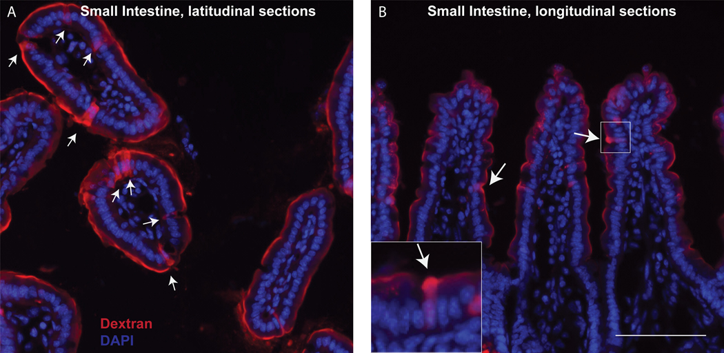
Figure 2:
sections of small intestine in longitudinal sections (left) and latitudinal sections (right) following GAP labeling (dextran, red) with optional WGA staining (WGA, green), as described in section 4 step H. White arrows denote GAP: goblet cell containing dextran and WGA, green arrow denotes a WGA+ Goblet cell that is not forming a GAP and does not contain dextran. Scale bar = 100 μm

