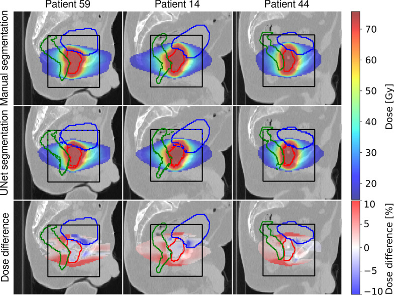
Fig. 3.
Dose distributions on sagittal slices of three test patients showing (left) the worst, (middle) the average and (right) the best agreement quantified by the gamma-index of the treatment plan optimized on (top) the manual segmentation and (middle) the 3D U-Net segmented images. Additionally, relative dose differences are presented. For improved visibility, dose below 25% of the dose prescribed to PTV and deviations below 0.4% on the difference plot are not displayed. Ground truth contours of (green) rectum, (blue) bladder and (red) prostate are also shown. The black box indicates the region where the contours were predicted by the U-Net

