
Figure 4. CR and CB labeling in caudal midline thalamus.
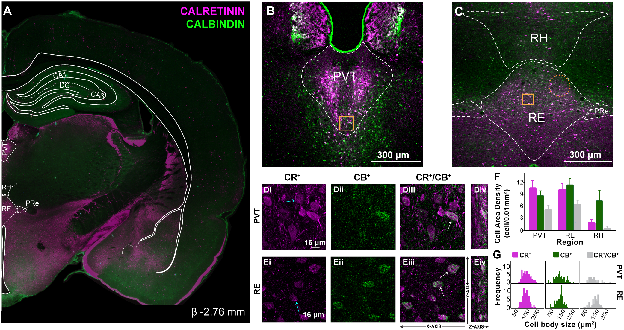
A: Representative coronal section (β −2.76 mm) showing immunofluorescent localization of CR+ and CB+ cell and fiber densities in caudal midline thalamus. Overlay shown adapted from Swanson (2018) to highlight midline thalamic structures. CR shown in magenta, CB in green.
B: Confocal image demonstrating distribution of CR+ and CB+ cells in PVT and PT caudally. In PVT, CR+ cells clustered mediolaterally and CB+ cells were predominantly seen in PVT ventral borders.
C: In RE, CB+ cells were prominent in dorsal and lateral borders, while CR+ cells were observed more mediolaterally, often overlapping with CB+ cells. CR+ fibers continued to be abundant ventrally (above 3V). Gold squares represent region of 60X magnification shown in inset. Orange dotted circles indicate regions in which there is sparse or no expression of calcium binding cells. Scale bar = 300μm.
D: Confocal images illustrating CR+ (Di), CB+ (Dii) and dual labeled CR+/CB+ (Diii) immunoreacted cell bodies in PVT. As in previous levels, three cell types are visualized: CR+ only cells (Di, blue arrow), CB+ only cells (not shown) and dual CR+/CB+ cell bodies (Diii, white arrows). The Z-axis from these optical sections are shown to the right at a 70° angle for visualization purposes (Div).
E: Confocal images in RE (Ei-Eiv). Scale bar = 16μm.
F: Comparison of CR+ and CB+ cell area density in PVT, RE and RH in caudal levels of the midline thalamus (cells/0.01mm2). Error bars represent SEM.
G: Frequency distribution of CR+, CB+ and CR+/CB+ immunoreacted cell body size (μm2) in caudal PVT and RE.
Abbreviations: β, bregma; CA1, CA1 subfield of the hippocampus; CA3, CA3 subfield of the hippocampus; CB, calbindin; CR, calretinin; DG, dentate gyrus; PVT, paraventricular; PRe, perireuniens; RE, nucleus reuniens, RH, rhomboid, SEM, standard error of the mean.