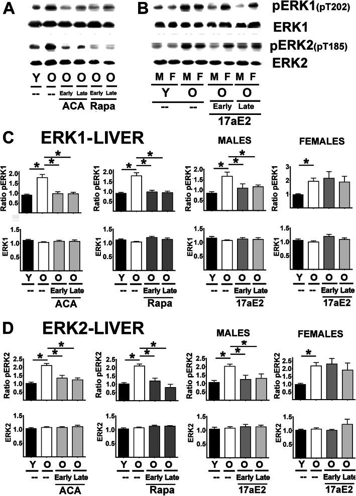
Fig. 3.
Effect of ACA, Rapa and 17aE2 treatments on liver ERK1 and ERK2 signaling in liver. A Representative western blots for phospho-ERK1 at Threonine 202 (pERK1), phosphor-ERK2 at Threonine 185 (pERK2) and total levels of ERK and ERK2 protein in liver samples from young untreated (Y), old untreated (O), old treated with ACA or Rapa (O, early and late treatments). B Same as A with 17aE2 treatment separated by sex (males = M, females = F). C) Bar graphs represent the mean ± SEM of pERK1 ratios and ERK1 protein levels in liver samples obtained from 16 young and 16 old mice plus, at least, 8 mice for each of the ACA and Rapa treatments groups. Data for 17aE2 was obtained from 8 young males and 8 young females, 8 old males and 8 old female mice, while treatments represent a minimum of 4 mice for each treatment and sex group. All values have been normalized to young control females as described in the Methods section. The (*) indicates statistical significance (p < 0.05) in a t-test between the indicated pair of groups. D Bar graphs represent the mean ± SEM for pERK2 ratios and ERK2 protein levels in kidney samples as described above

