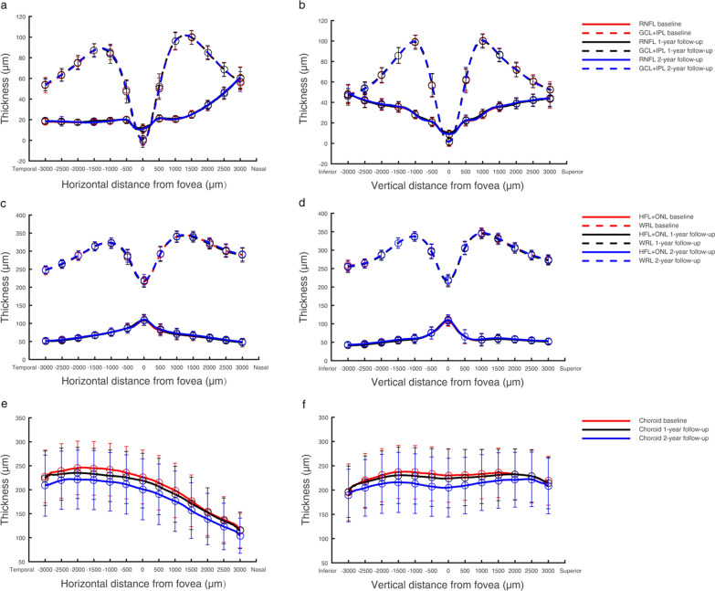
Fig. 2.
Thickness profiles of the retina and choroid in 168 myopic children. The thickness profiles of three intra-retinal layers, the total retina, and the choroid in horizontal and vertical scans were obtained by SD-OCT and averaged over each test. Thickness values for the RNFL, GCL + IPL, HFL + ONL, and WRL were similar; therefore, they overlapped each other in the figure and cannot be seen separately. a, b Horizontal (left) and vertical (right) thickness profiles of the RNFL and GCL + IPL; c, d horizontal (left) and vertical (right) thickness profiles of the HFL + ONL and whole retina; e, f horizontal (left) and vertical (right) thickness profiles of the choroid. RNFL, retinal nerve fibre layer; GCL + IPL, ganglion cell layer and inner plexiform layer; HFL + ONL, Henle fibre layer and outer nuclear layer; WRL, whole retinal layer

