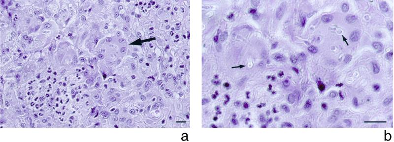
FIG. 1.
Hematoxylin-eosin stain of a biopsy specimen from the lesion of the right thumb. (a) Margin of an abscess with accumulation of polymorphonuclear leukocytes (left lower corner) and numerous multinucleated giant cells (arrow) engulfing fungal elements. (b) The fungal elements are surrounded by hyaline capsules (arrows) which become visible by lowering the condensor diaphragm. Bar, 10 μm.

