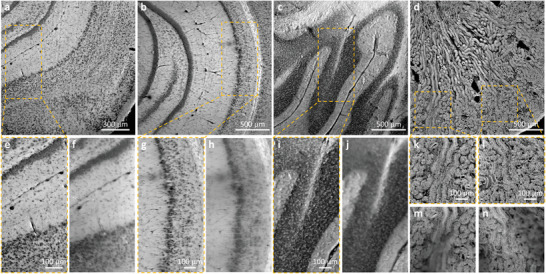
Figure 3.

CHAMP imaging of thick and unprocessed mouse brain/kidney tissues. a–c) CHAMP images of fixed and unprocessed mouse brain tissues hand‐cut at different coronal planes with thickness ∼ 5 mm. d) CHAMP image of a fixed and unprocessed mouse kidney tissue sectioned with thickness ∼200 µm. e,g,i,k,l) Zoomed‐in CHAMP images of orange dashed regions in (a–d). f,h,j,m,n) The corresponding wide‐field images captured with a 0.3‐NA imaging objective with uniform illumination.
