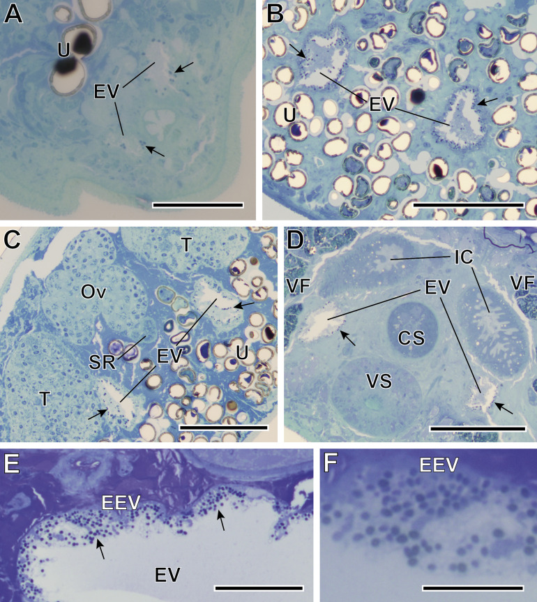
Figure 2.
Semithin sections of Bacciger israelensis stained with methylene-blue at different levels showing the presence of many spores (black arrows). (A–B) Sections at 35 and 155 μm from the posterior extremity of the digenean showing the two arms of the Y-shaped excretory vesicle (EV); (C) Section at 210 μm from the posterior extremity at the level of genitalia, showing both testes (T) and the characteristic trilobed ovary (Ov); (D) Section at 310 μm from the posterior extremity at the level of the ventral sucker (VS) and the cirrus sac (CS); (E) Detail of the excretory vesicle at 150 μm from the anterior extremity showing numerous T. baccigeri n. gen., n. sp. in its epithelium (EEV); (F) Enlarged detail of the epithelium of the excretory vesicle. IC, intestinal caeca; SR, seminal receptacle; U, uterus; VF, vitelline follicles. Scale bars: A, E = 50 μm; B–D = 100 μm; F = 20 μm.

