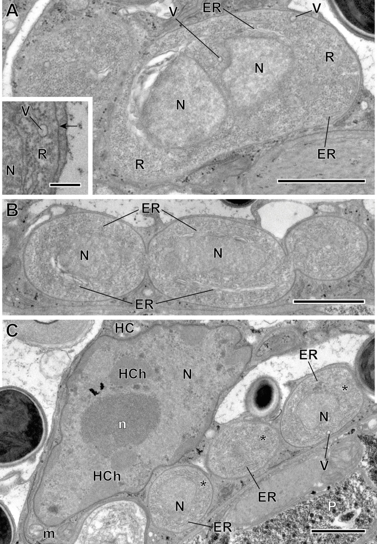
Figure 4.
Merogonic stages of Toguebayea baccigeri n. gen., n. sp. (A–B) Dividing meronts; (C) Merozoites (*) in a host cell (HC); (inset) Detail at high magnification of the meront plasma membrane (black arrow). ER, endoplasmic reticulum; HCh, heterochromatin cluster; m, mitochondrion; N, nucleus; n, nucleolus; P, parenchyma; R, ribosomes; V, vacuoles. Scale bars: A–C = 1 μm; inset = 0.2 μm.

