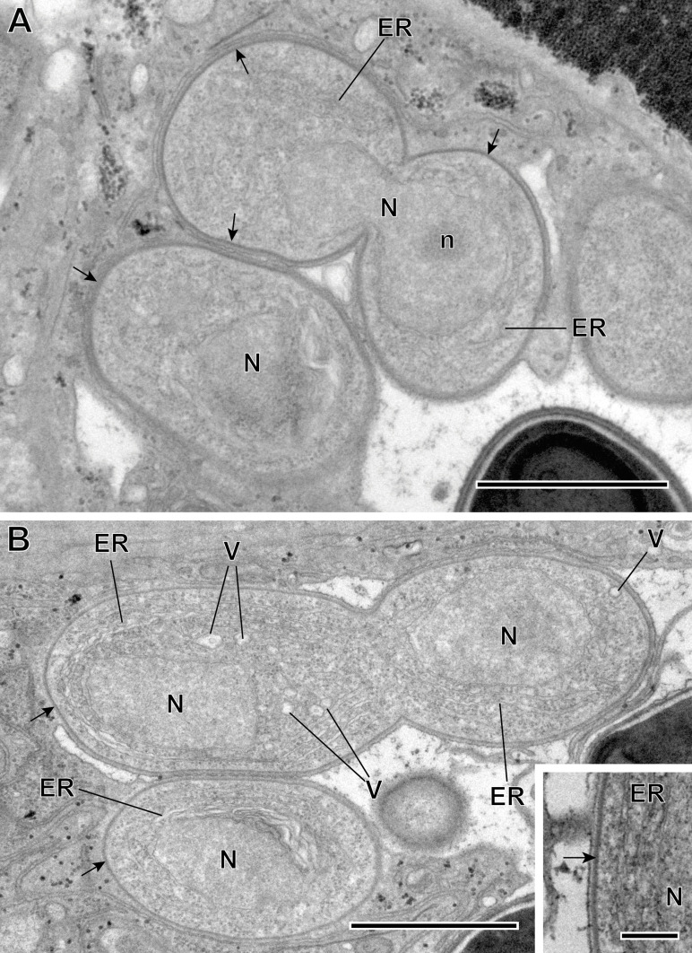
Figure 5.
(A–B) Sporogonic stage of Toguebayea baccigeri n. gen., n. sp. Note the thick membrane of sporonts (black arrows) in comparison with meronts; (inset) Detail at high magnification of the sporont plasma membrane (black arrow). ER, endoplasmic reticulum; N, nucleus; n, nucleolus; V, vacuoles. Scale bars: A–B = 1 μm; inset = 0.2 μm.

