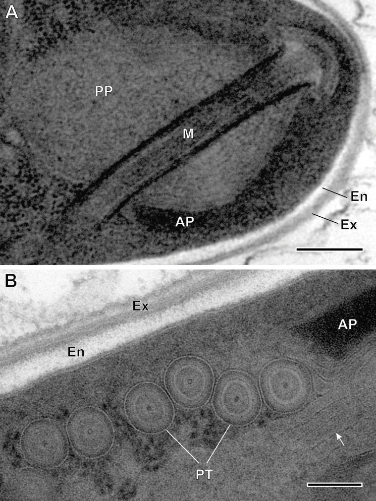
Figure 9.
Ultrastructural details of mature spores of Toguebayea baccigeri n. gen., n. sp. (A) Detail of the apical part showing the manubrium (M); (B) Detail showing cross-sections of the polar tube (PT) composed of several concentric electron-dense and electron lucent layers surrounding a central tubular structure. Note the lamellar external area (white arrows) of the posterior polaroplast (PP). AP, anterior polaroplast; En, endospore; Ex, exospore. Scale bars: A = 0.2 μm; B = 0.1 μm.

