FIG 3.
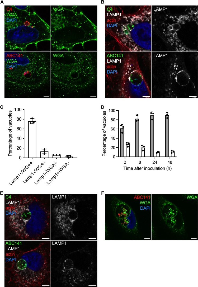
Multiplication of A. baumannii C4 and ABC141 occurs in large vacuoles positive for LAMP1. Human cells were infected with A. baumannii C4 or ABC141, immunolabeled 24 hpi, and analyzed using confocal immunofluorescence microscopy. Representative images are shown. Scale bars correspond to 5 μm. (A) Membranes of A549 cells were labeled with WGA (green), the nucleus with DAPI (blue), and A. baumannii with antibodies (red). A. baumannii C4 and ABC141 multiply in Acinetobacter-containing vacuoles (ACVs). (B) A549 cells were labeled with anti-LAMP1 antibodies (gray) with phalloidin and DAPI to visualize actin cytoskeleton and nucleus, respectively, and A. baumannii isolates were labeled with specific antibodies (green). ACVs are positive for LAMP1 in A549 cells. (C) The numbers of ACVs positive for LAMP-1 and/or WGA staining were counted at 24 hpi for cells infected by A. baumannii ABC141. ACVs positive for both LAMP1 and WGA staining represent 78% of intracellular bacteria clusters. Data correspond to mean ± SD from 3 independent experiments. (D) A549 cells infected by A. baumannii ABC141 were labeled with Lysotracker DND-99 (white bars) or LAMP1 (gray bars) at 2, 8, 24, and 48 h postinfection to visualize acidic compartments and the kinetics of acquisition of the late endosomal marker LAMP1. The majority of ACVs do not show features of acidic lysosomes. Data correspond to mean ± SD from 3 independent experiments. (E) EA.hy 926 cells were labeled with anti-LAMP1 antibody (gray), anti-A. baumannii specific antibodies (green), and with phalloidin and DAPI to visualize actin cytoskeleton and nucleus. ACV are positive for LAMP1 in EA.hy 926 cells. (F) Representative images of human primary keratinocytes infected with A. baumannii ABC141 for 24 h. Nuclei were labeled with DAPI (blue), ABC141 were labeled with a specific antibody (red), and membranes with WGA (green).

