FIG 4.
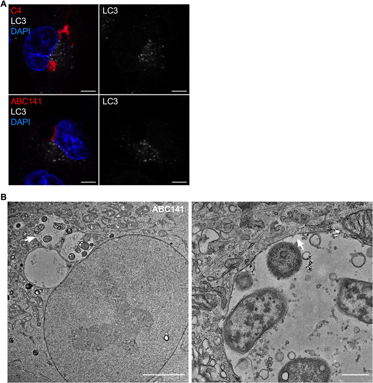
ACVs are single-membrane vacuoles that do not colocalize with the autophagy marker LC3. (A) A549 cells were infected with A. baumannii C4 or ABC141, immunolabeled 24 h postinfection, and analyzed using confocal immunofluorescence microscopy. A. baumannii (red) and LC3 (white) were labeled with antibodies and nucleus with DAPI (blue). ACVs do not colocalize with LC3. Representative images are shown. Scale bars correspond to 5 μm. (B) Transmission electron microscopy of infected A549 cells by A. baumannii ABC141 24 h postinfection. The scale bar corresponds to 5 μm. The second picture represents a zoom of the vacuole indicated by the white arrow. The scale bar corresponds to 500 nm. A. baumannii ABC141 multiplies in ACVs with a single membrane.

