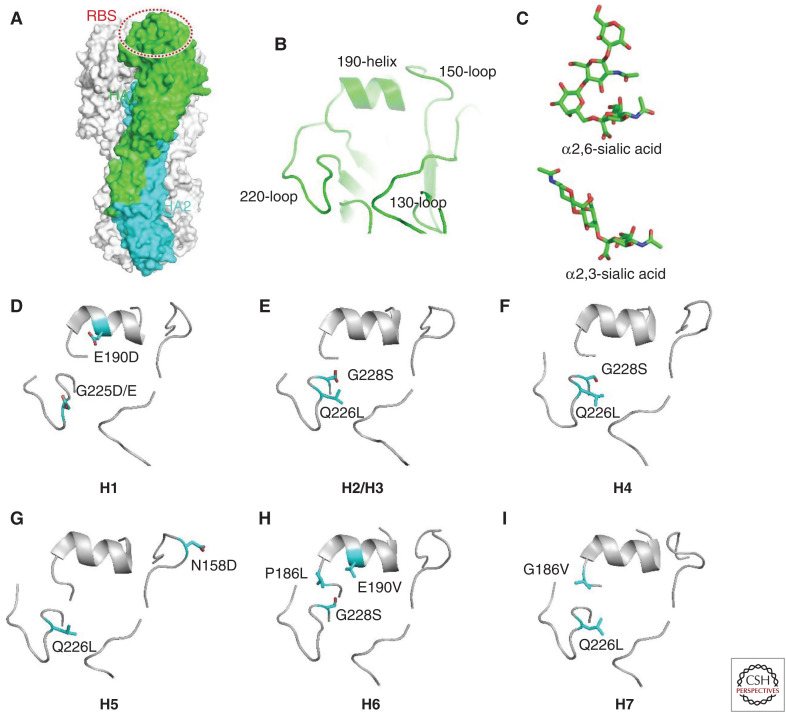
Figure 4.
Molecular determinants for receptor binding properties of different hemagglutinin (HA) subtypes. (A) Structure of HA trimer. One of the protomers is colored by subunits with HA1 in green and HA2 in cyan. The receptor binding site (RBS) is indicated by a dashed oval. (B) Close-up view of the structural motifs within RBS. (C) Structures of different sialic acid receptors. (D–I) The key determinant residues (sticks) for the receptor binding properties of different HA subtypes.

