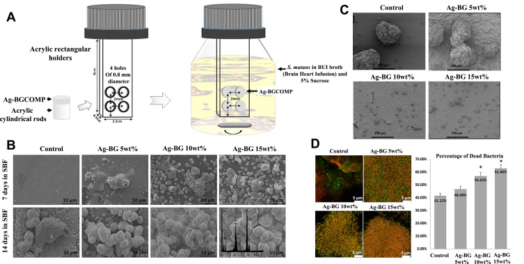
Figure 3.
The AgBM was incorporated into resin composite. (A) Schematic illustration of biofilm formation by S. mutans on the surface of AgBM. (B) SEM images of the surface morphology of the AgBM specimens after immersion. (C) SEM images of the biofilm formed on the surface of the specimens. (D) CLSM representative images of the live and dead bacteria in the biofilms formed on the surface of the AgBM. *Shows the statistical significant difference . Reproduced with permission from Chatzistavrou X, Lefkelidou A, Papadopoulou L, et al. Bactericidal and Bioactive Dental Composites. Front Physiol. 2018;9:103. Copyright © 2018 Chatzistavrou, Lefkelidou, Papadopoulou, Pavlidou, Paraskevopoulos, Fenno, Flannagan, González-Cabezas, Kotsanos and Papagerakis. This is an open-access article distributed under the terms of the Creative Commons Attribution License (CC BY).61

