
Figure 1.
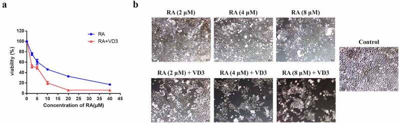
Cell viability of the HeLa cells. (a) Following treatment with RA (2.5, 5, 10, 20 and 40 μM/L) and RA combined with 100 nM/L VD3 for 48 h, the viability of HeLa cells was determined using MTT assay. Data was presented as mean ± SD of three independent experiments. P < 0.01 versus the control groups (0 μM/L RA and 0 μM/L VD3). (b) HeLa cells under microscope. HeLa cells were cultured for 48 h with different concentrations of RA (2, 4 and 8 μM/L) and RA combined with 100 nM/L VD3. The morphological changes were investigated under microscope. (×100). RA: Raddeanin A; RA+VD3: Raddeanin A combined with 100 nM/L VD3.