
Figure 1.
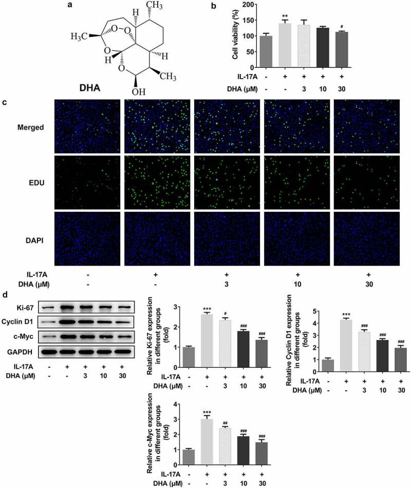
DHA inhibits over proliferation of IL-17A-stimulated HaCaT cells. (a) The chemical structure of DHA; (b) Cell viability following the different concentration DHA treatment was detected by CCK-8; (c) Cell proliferation was detected with EDU staining; (d) Cell proliferation-related protein expressions were analyzed by Western blotting, including Ki-67, cyclin D1 and c-Myc. **P < 0.01 and ***P < 0.001 vs. control. #P < 0.05, ##P < 0.01 and ###P < 0.001 vs. IL-17A. DHA, dihydroartemisinin.