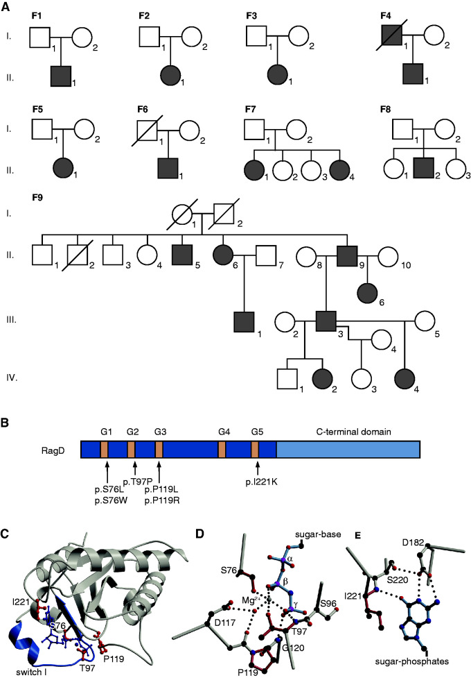
Figure 2.
Mutations in the RagD protein are located in the GTP-binding sites. (A) Pedigrees of all families included in this study. Filled symbols represent affected individuals. Squares indicate male family members and circles represent female family members. (B) Domain organization of RagD with GTP-binding motifs in dark blue (G1–G5). Mutations are indicated with an arrow. (C) Crystal structure of RagD in complex with a GTP analogue (GppNHp; Protein Data Bank entry 2q3f). Mutated residues are shown in red. GppNHp and the coordinated Mg2+ are depicted in dark blue. (D and E) Detailed view of the (D) phosphate moiety of the nucleotide and (E) the nucleotide base. Affected residues are colored in light red. Dotted lines indicate hydrogen bonds.

