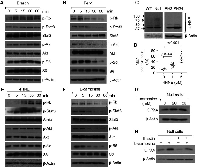
Figure 6.
Pkd1 mutant renal epithelial cell proliferation was regulated by 4-HNE produced during ferroptotic process. (A and B) Western blot analysis of the expression and phosphorylation of Rb, STAT3, Akt, and S6 in Pkd1 null MEK cells treated with erastin (10 μM) (A) or Fer-1 (1 μM) (B) at indicated time points. (C) Western blot analysis of the expression of 4HNE in Pkd1 WT and null MEK cells (left panel) and PH2, PN24 cells (right panel). (D) Ki67 staining indicated that cell proliferation was increased in Pkd1 null MEK cells treated with 4HNE compared with those cells treated with vehicle. The percentage of Ki67-positive nuclei of Pkd1 null MEK cells was quantified from an average of 1000 nuclei per field. Statistical data are presented as the mean±SEM. (E) Western blot analysis of the expression and phosphorylation of Rb, STAT3, Akt, and S6 in Pkd1 null MEK treated with 4HNE (5 μM) at indicated time points. (F) Western blot analysis of the expression and phosphorylation of Rb, STAT3, Akt, and S6 in Pkd1 null MEK cells treated with L-carnosine at indicated time points. (G and H) Western blot analysis of the expression of GPX4 in Pkd1 null MEK cells treated with L-carnosine at indicated concentrations (G) and treated with erastin with or without L-carnosine (H).

