Figure 7.
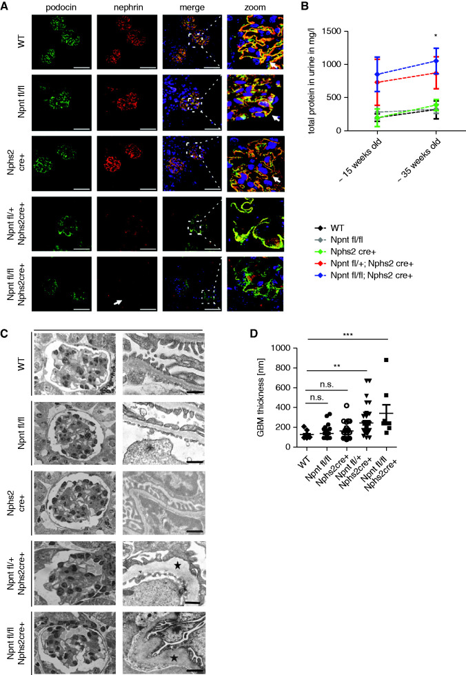
Podocyte-specific Npnt knockout mice show altered expression of podocyte markers and structural impairments of the GBM. (A) Immunofluorescence staining for nephrin and podocin in WT mice, Npnt fl/fl mice, Nphs2 cre+ mice, Npnt fl/+; Nphs2 cre+ mice and Npnt fl/fl; Nphs2 cre+ mice. Nuclei were stained with Hoechst. The merged pictures and higher magnifications of merged pictures are given to illustrate colocalization of podocin and nephrin (white arrows in zoom picture). In Npnt fl/fl; Nphs2 cre+ mice nephrin expression is reduced (white arrow). Scale bar=50 µm. (B) Proteinuria in mg/L in WT, Npnt fl/fl, Nphs2 Cre+, Npnt fl/+; Nphs2 cre+ and Npnt fl/fl; Nphs2 cre+ mice at approximately 15 and approximately 35 weeks of age. *P<0.05. (C) Semithin sections of glomeruli and TEM picture of the ultrafiltration barrier of 15-week-old mice. WT, Npnt fl/fl; Nphs2 cre+, Npnt fl/+; Nphs2 cre+; and Npnt fl/fl; Nphs2 cre+ mice are shown as indicated. Black star illustrates GBM pathology. Scale bar=500 nm. (D) Statistical analysis of GBM width of 15 weeks old WT, Npnt fl/fl, Nphs2 cre+, Npnt fl/+; Nphs2 cre+ and Npnt fl/fl; Nphs2 cre+ mice. *P<0.05, **P<0.01, ***P<0.001. WT, wild type.

