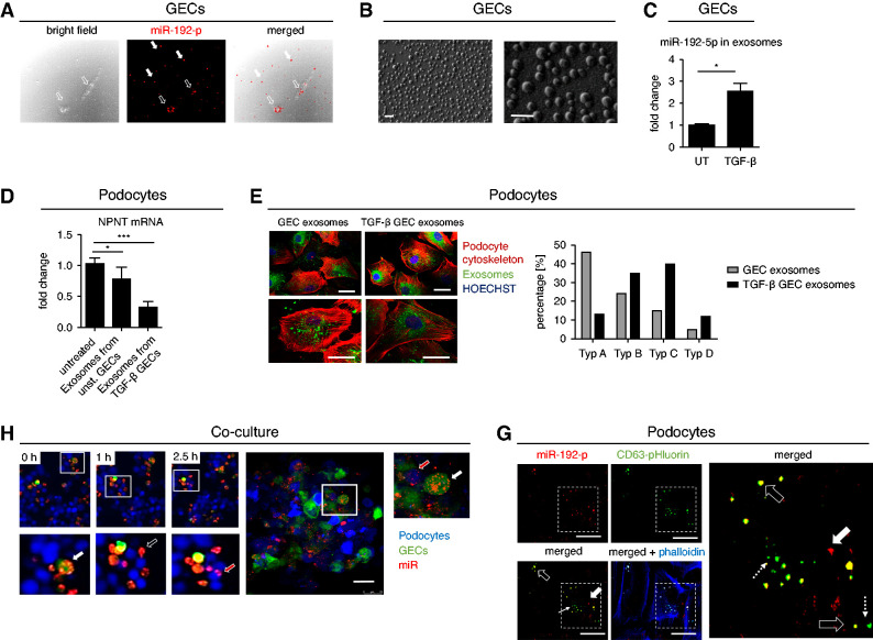
Figure 8.
GEC-derived exosomes carry miR-192-5p that is taken up by podocytes causing a decrease in NPNT expression. (A) Secretion of Alexa555-tagged miR-192-5p mimic (red) from GECs. White arrows show miR-192-5p outside the cells, black arrow heads show miR-192-5p inside the GECs. Scale bar = 50 µm. (B) Surface electron microscopy image of GEC-derived exosomes secreted in the cell culture supernatant. Scale bar 20 nm. (C) qPCR for miR-192–5p expression normalized to U6 RNA in secreted exosomes derived from unstimulated and TGF-β-stimulated GECs. Change in miR expression is given as fold change compared with the unstimulated condition. n=4; *P<0.05. (D) qPCR for NPNT mRNA in cultured human podocytes treated with exosomes derived from untreated and TGF-β–treated GECs. n=3, ***P<0.001. (E) Treatment of cultured human podocytes with exosomes (labeled in green) derived from unstimulated and TGF-β–stimulated GECs. Exosomes derived from TGF-β–stimulated GECs caused cytoskeletal rearrangements (labeled in red). Scale bar 25 µm. (F) Left: Still frames of a time-lapse of three-dimensional coculture of GFP-labeled human GECs (green) with podocytes that were in vivo labeled with cell proliferation dye eFluor 450 (blue). GECs were transfected with Alexa555-tagged miR-192-5p mimic (red) before coculture. Higher magnifications in the lower row illustrate miR-192-5p in GECs (white arrow), miR-192-5p outside the cells (black arrow) and miR-192-5p inside podocytes (red arrow). Right: Confocal fluorescent microscopy picture of the same coculture after 24 hours to show higher resolution. Red fluorescent miR-192-5p is located inside the green fluorescent GECs (white arrow) and inside the blue fluorescent podocytes (red arrow). Scale bar = 50 µm. (G) Endocytosis of miR-192-5p containing GEC-derived exosomes in cultured human podocytes. CD63-pHluorin plasmid was electroporated into GECs before transfection with Alexa555-tagged miR-192-5p. Exosomes were isolated from GECs and incubated with human podocytes. Black arrows show overlap of red fluorescent miR-192-5p and green fluorescent exosomes (miR-192-5p containing exosomes), dotted white arrows show exosomes without miR-192-5p and thick arrow shows naked miR-192-5p. Scale bar = 50 µm.

