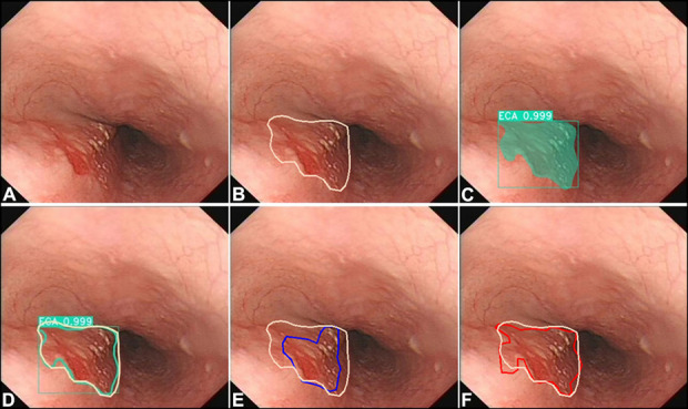
Figure 3.

Comparing the delineation performance of the model with that of endoscopists. (a) A case of cancer in the esophagus with white light imaging. (b) Margins of the same lesion under WLI were manually delineated (white polygonal frames, used as the gold standard) by an expert who took the margins of the lesions under NBI, iodine staining, and resection specimen as reference. (c) The AI model correctly detected the lesion by indicating it with a square frame and a polygonal frame (dark cyan). (d) Margins of the same lesion under WLI were delineated AI model (a dark cyan polygonal frame) and the gold standard (white polygonal frames). (e) Margins of the same lesion under WLI were delineated by a senior endoscopist (a blue polygonal frame) and the gold standard (white polygonal frames). (f) Margins of the same lesion under WLI were delineated by an expert endoscopist (a red polygonal frame) and the gold standard (white polygonal frames). AI, artificial intelligence; NBI, narrow-band imaging; WLI, white light imaging.
