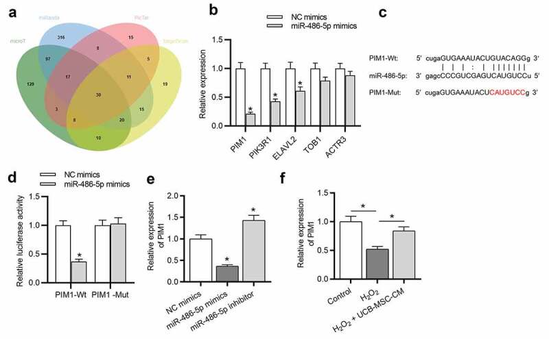
Figure 4.

PIM1 is targeted by miR-486-5p. (a) Bioinformatics tools (microT, miRanda, PicTar and TargetScan) were used and Venn diagram exhibits 30 potential targets of miR-486-5p. (b) After miR-486-5p overexpression, the expression of targets was measured by RT-qPCR. (c) MiR-486-5p binding site in the wild type or mutant sequence of PIM1-3ʹUTR. (d) The binding capacity between miR-486-5p and PIM1 was confirmed by a luciferase reporter assay. (e) The expression of PIM1 in H2O2-treated cells transfected with miR-486-5p mimics or inhibitor was measured by RT-qPCR. (f) RT-qPCR was used to measure the expression of PIM1 in L02 cells after H2O2 stimulation and UCB-MSC-CM treatment. *P < 0.05
