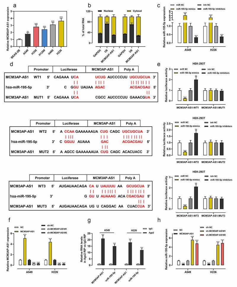
Figure 2.

MCM3AP-AS1 was specifically regulated by miR-195-5p
(a) MCM3AP-AS1 expressions in BEAS-2B cells and NSCLC cells were measured by qRT-PCR. (b) Subcellular localization of MCM3AP-AS1 in A549 and H226 was assessed by qRT-PCR after nuclear–cytoplasm fractionation. (c) MiR-195-5p mimics or miR-195-5p inhibitors were transfected into A549 and H226 cells, respectively, and the transfection efficiency was examined by qRT-PCR. (d) The schematic map of the MCM3AP-AS1 WT and MCM3AP-AS1 MUT binding sites for miR-195-5p, which was predicted by LncBase Predicted v.2 (Score: 0.947). (e) MCM3AP-AS1-WT (WT1, WT2 and WT3) or MCM3AP-AS1-MUT (MUT1, MUT2 and MUT3) was co-transfected into HEK-293 T cells with miR-195-5p mimics or miR-195-5p inhibitors, and the relative luciferase activity was measured. (f) Transfection efficiency of MCM3AP-AS1 overexpression plasmids, sh-MCM3AP-AS1#1 or sh-MCM3AP-AS1#2 was detected by qRT-PCR. (g) The interaction between MCM3AP-AS1 and miR-195-5p in A549 and H226 cells was analyzed by RIP experiment. (h) Effect of MCM3AP-AS1 knockdown and overexpression on miR-195-5p expression in A549 and H226 cells was detected by qRT-PCR. All of the experiments were performed in triplicate. ** P < 0.01 and *** P < 0.001.
