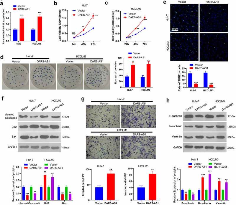
Figure 2.

DARS-AS1 subserved HCC proliferation, invasion and EMT
A. DARS-AS1 overexpression models were constructed in Huh-7 and HCC-LM3, respectively, and the DARS-AS1 expression was tested by RT-qPCR. B and C. CCK-8 experiment was implemented to verify Huh-7 and HCC-LM3 cell proliferation. D. Cell proliferation was examined by the colony formation assay. E. Cell apoptosis was monitored by the TUNEL assay. F. The expression of Caspase3, Bax and Bcl2 in HCC cells was compared by WB. G. Transwell assay was performed to test cell invasion. H. The expression of E-cadherin, N-cadherin and Vimentin in HCC cells was compared by WB. NS, **, *** representedP> 0.05,P< 0.01,P< 0.001, compared with the Vector group. N = 3.
