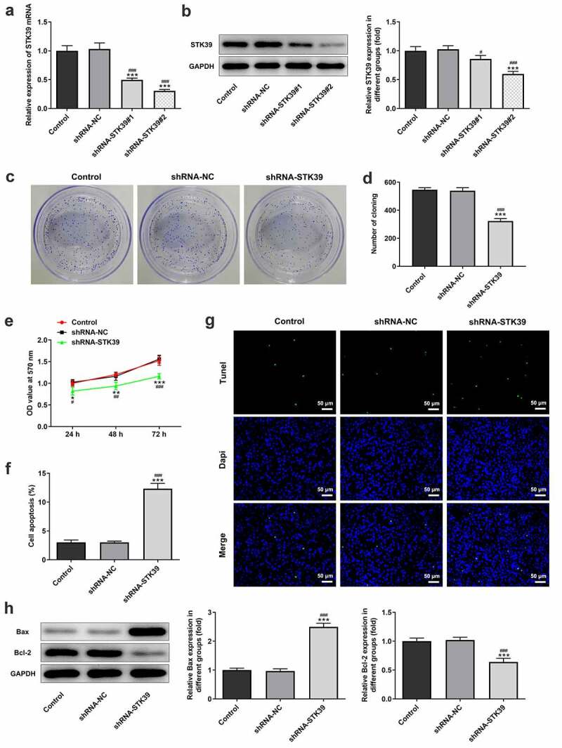
Figure 2.

STK39 knockdown inhibited proliferation and induces apoptosis of Hep3b cells
(a,b) The mRNA and protein expression of STK39 in control Hep3b cells or cells that transfected with indicated shRNAs was determined with RT-qPCR and western blotting. ***P < 0.001 vs Control; #P < 0.05 and ###P < 0.001 vs shRNA-NC. (c,d) Colony formation assay was performed to observe cell proliferation. (e) Cell viability at 24, 48 and 72 h was measured respectively by means of MTT assay. (f,g) Cell apoptosis was observed using Tunel staining (×200); (h) The protein expression of Bax and Bcl-2 was detected via western blot assay. *P < 0.05, **P < 0.01, ***P < 0.001 vs Control; #P < 0.05, ##P < 0.01 and ###P < 0.001 vs shRNA-NC.
