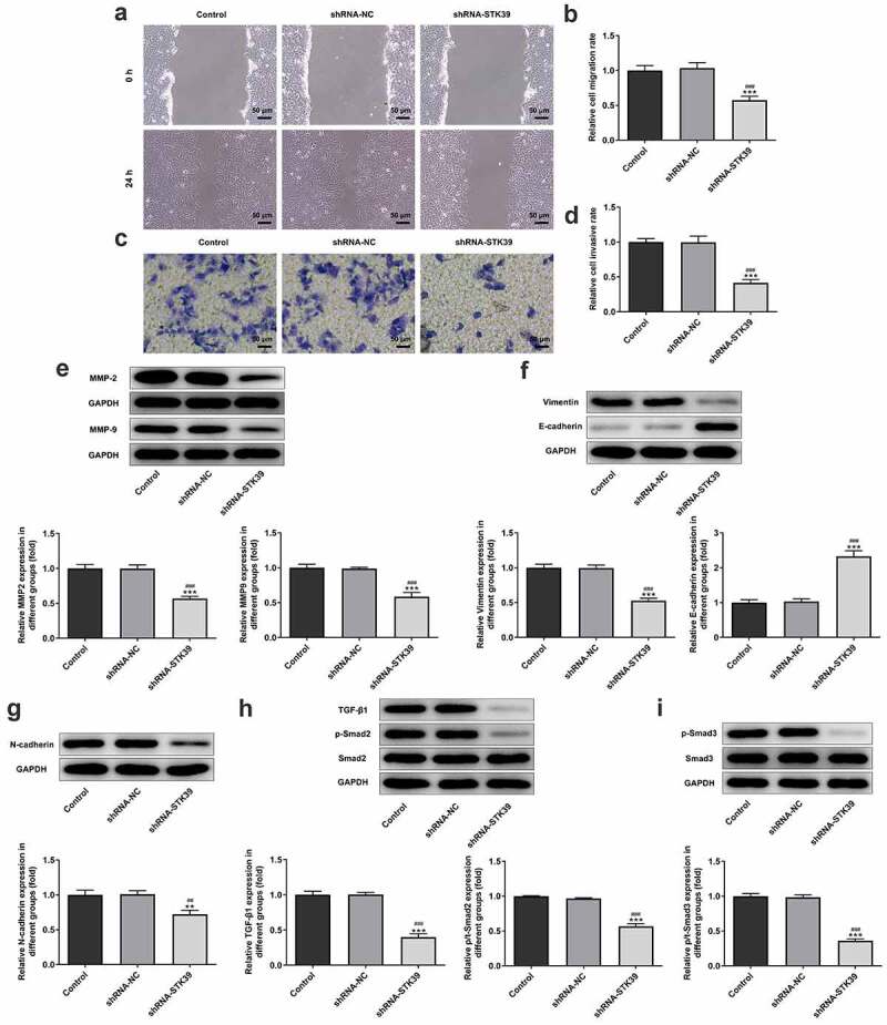
Figure 3.

STK39 knockdown suppressed migration, invasion, EMT and TGF-β1/Smad2/3 signaling of Hep3b cells
(a,b) Hep3b cells were transfected with shRNA-NC or shRNA-STK39 or not, then cell migration was assessed using wound healing assays. (c-d) Cell invasion was evaluated with transwell assay. (e) The protein expression of MMP2 and MMP9 was detected using western botting. (f,g) The expression of EMT-related proteins including vimentin, E-cadherin and N-cadherin was assessed by means of western blot assay. (h) TGF-β1, p-Smad2/Smad2 and p-Smad3/Smad3 expression was detected using western blot. **P < 0.01 and ***P < 0.001 vs Control; ##P < 0.01 and ###P < 0.001 shRNA-NC.
