
Figure 11.
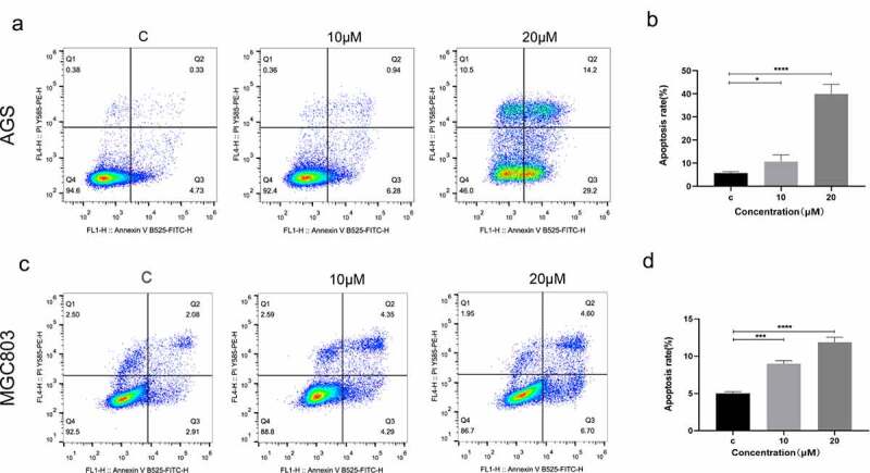
(e)-SIS3 induces apoptosis of gastric cancer cells
Flow cytometry results for apoptosis [apoptosis ratio was calculated as (Q2+ Q3)/(Q1+ Q2 + Q3+ Q4)] of AGS (A and B) and MGC803 (C and D) cells incubated with 10 μM and 20 μM (E)-SIS3 or an equal volume of DMEM medium for 24 hours. (E)-SIS3 significantly induced apoptosis in AGS and MGC803 cells. (E)-SIS3 induced apoptosis in a concentration-dependent manner. Data are presented as mean ± SD, N = 5; *P < 0.05; ***P < 0.001; ****P < 0.0001, compared with control.