
Figure 4.
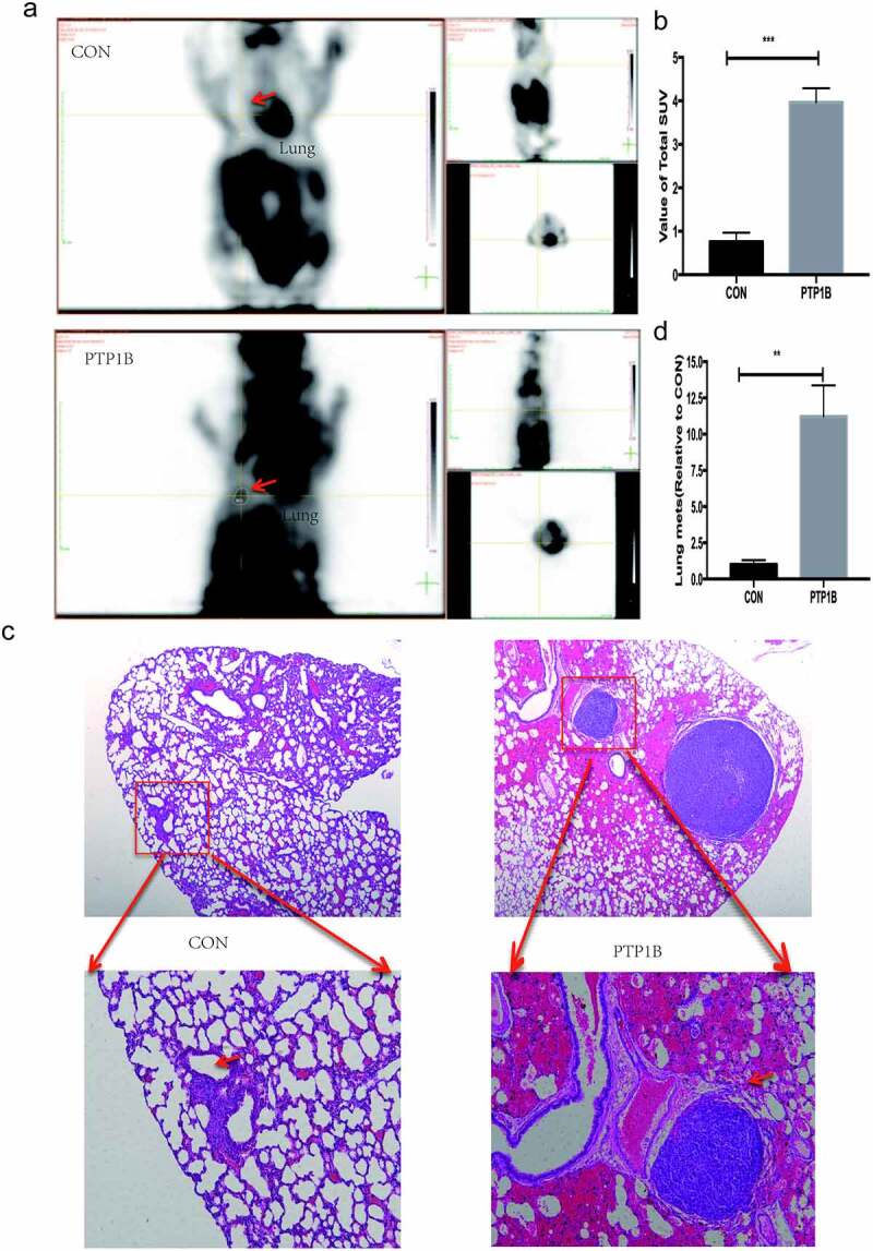
PTP1B promotes tumor metastasis in vivo
A. PET-CT images showing the lung metastasis area (red arrow).B. Analysis of the SUV value in the lung metastases.C. Images of lung metastases subjected to HE staining. Original magnification, 40× and 200 × .D. Analysis of the relative lung metastasis based on the number and area of metastases.