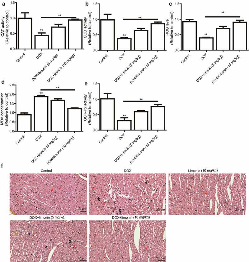
Figure 3.

Limonin ameliorates DOX-induced cardiac injury in rat. (a) CAT activity was detected in heart tissues from rat treated differently as indicated. (b) SOD activity was examined in heart tissues from rat treated differently as indicated. (c) ROS level was determined in heart tissues from rat treated differently as indicated. (d) MDA activity was evaluated in heart tissues from rat treated differently as indicated. (e) GSH-Px activity was measured in heart tissues from rat treated differently as indicated. **p < 0 01 vs control. (f) Myocardial cell injury was detected heart tissues from rat treated differently as indicated. Control group: showing branched striated muscle fibers with central vesicular nuclei and light eosinophilic cytoplasm. Some blood capillaries present in between fibers. DOX group: showing area of degenerated wavy muscle fibers with absent striations (black arrows). Some fibers appear with pyknotic nuclei (red arrows), others with absent nuclei (*). Notice marked congested blood capillaries with inflammatory cells infiltration (&) and hemosiderin pigment (#) between muscle fibers. Limonin group: showing branched striated muscle fibers with central vesicular nuclei and light eosinophilic cytoplasm. Some blood capillaries present in between fibers. DOX + limonin (5 mg/kg): showing some areas of degenerated muscle fibers with absent striations (black arrows), while others are showing preserved muscle fiber with central oval nuclei (red arrows). Notice marked scattered dilated congested capillaries (c) with cloudy fatty degeneration and intermuscular edema (#). DOX + limonin (10 mg/kg) group: showing areas of preserved muscle fiber striations with central oval nuclei (red arrows). Notice some dilated congested capillaries (c) with minimal areas of edema between fibers. (HE× 200, scale bar = 20 µm)
