Fig. 2.

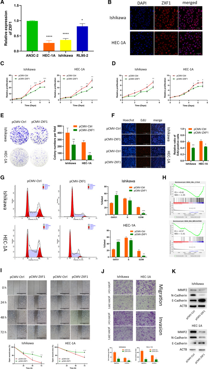
LncRNA‐ZXF1 overexpression reduces the viability and invasion of EEC cells. (A) Comparison of ZXF1 expression in the EEC cell lines using qRT‐PCR. (B) Fluorescence in situ hybridization of ZXF1 was performed in EEC cell lines. ZXF1 was detected in both the cytoplasm and nucleus. Scale bars, 50 μm. (C) The Celltiter‐Glo curves showed that ZXF1 reduced the viability of EECs. Comparisons among multiple groups are analyzed using two‐way ANOVA. (D) The MTT assay showed that ZXF1 inhibited the proliferation of endometrial tumor cells. Comparisons among multiple groups are analyzed using two‐way ANOVA. (E) Colony formation experiments proved that ZXF1 reduces the number of colonies formed from single EEC cells. (F) DNA replication in the nucleus was detected using the EdU incorporation assay. After ZXF1 overexpression, DNA replication was inhibited. Scale bars, 100 μm. (G) Flow cytometry showed that ZXF1 arrested the cell cycle in Ishikawa and HEC‐1A cells. (H) All samples were divided into two groups according to the expression of ZXF1 in TCGA, and then, a GSEA was performed to enrich the cell cycle and DNA replication results. (I) Overexpression of ZXF1 in tumor cells used in the wound healing experiment inhibited migration. Scale bars, 100 μm. (J) Transwell migration and invasion assays were performed using Ishikawa and HEC‐1A cells transfected with pCMV‐ZXF1. The presence of ZXF1 weakened the migration and invasion capabilities of tumor cells. Scale bars, 100 μm. (K) Protein expression levels of invasion‐related markers in Ishikawa and HEC‐1A cells transfected with ZXF1 and control vectors. All data are mean ± SD. Significance calculated using the unpaired t‐test. *P < 0.05, **P < 0.01, ***P < 0.001,****P < 0.0001. Representative data are from three independent experiments.
