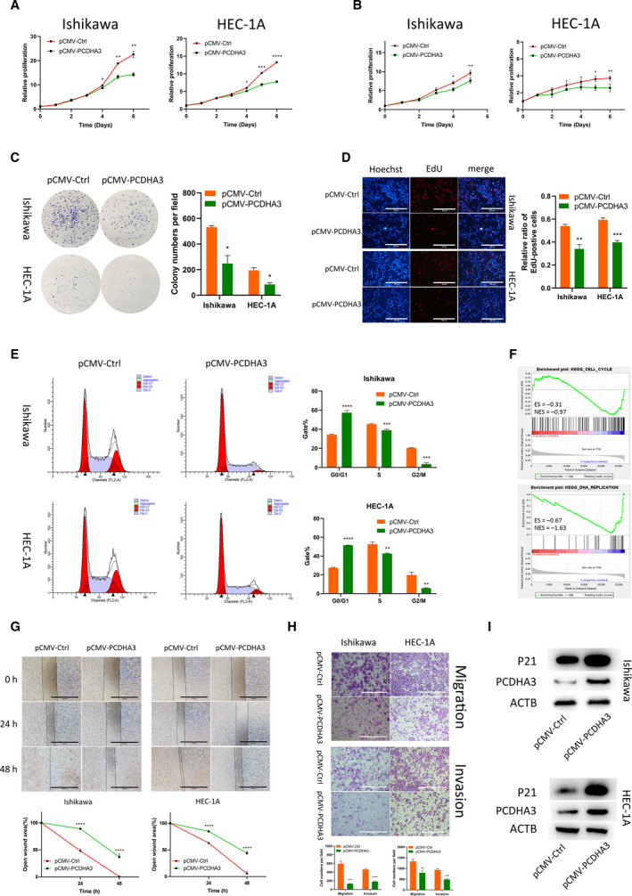
Fig. 5.

PCDHA3 inhibits the malignant phenotypes of endometrial cancer cells. (A) CellTiter activity was used to detect the proliferation of Ishikawa and HEC‐1A cells. The proliferation of EEC cell lines was inhibited after PCDHA3 overexpression. Comparisons among multiple groups are analyzed using two‐way ANOVA. (B) After pCMV‐PCDHA3 was transfected into endometrial cancer cells, MTT assays proved that PCDHA3 inhibited the proliferation of EEC cells. Comparisons among multiple groups are analyzed using two‐way ANOVA. (C). The amplification of single EEC cells transfected with pCMV‐PCDHA3 in colony formation experiments was suppressed. (D) PCDHA3 overexpression suppressed nuclear DNA replication in Ishikawa and HEC‐1A cells. Scale bars, 100 μm. (E) The cell cycle was detected using flow cytometry. PCDHA3 increased the proportion of EEC cells in G0/G1 phase and decreased the proportion of EEC cells in S and G2/M phase. (F) TCGA data were divided into two groups according to the median expression of PCDHA3. The GSEA using these two datasets showed that cell cycle and DNA replication were enriched. (G) Wound healing experiments were used to explore cell migration capabilities. Detection of the healing rate of the scratches at different time points showed that PCDHA3 prevented the healing of the scratch wounds. Scale bars, 100 μm. (H) The Transwell system was used to detect the migration and invasion capabilities of EEC cells. Compared with the control group, fewer cells transfected with pCMV‐PCDHA3 migrated and invaded. Scale bars, 100 μm. (I) PCDHA3 overexpression increased P21 protein expression in Ishikawa and HEC‐1A cells. All data are mean ± SD. Significance calculated using the unpaired t‐test. *P < 0.05, **P < 0.01, ***P < 0.001, ****P < 0.0001. Representative data are from three independent experiments.
