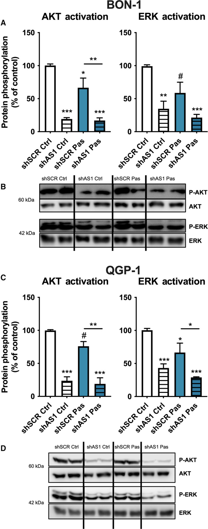
Fig. 4.

Silencing of SSTR5‐AS1 alters key SST5‐related signaling pathways in BON‐1 (A, B) and QGP‐1 (C, D) cells. Protein phosphorylation of AKT and ERK in both cell lines after SSTR5‐AS1 silencing (striped bars) and after 10 min of pasireotide treatment (Pas, blue). This activation was measured by western blot and normalized with total AKT/ERK. Asterisks (*P < 0.05; **P < 0.01; ***P < 0.01) indicate values that significantly differ between groups (one‐way ANOVA analysis); # symbol indicates values that significantly differ from control under t test. In all cases, data represent mean ± SEM of n = 4 independent experiments.
