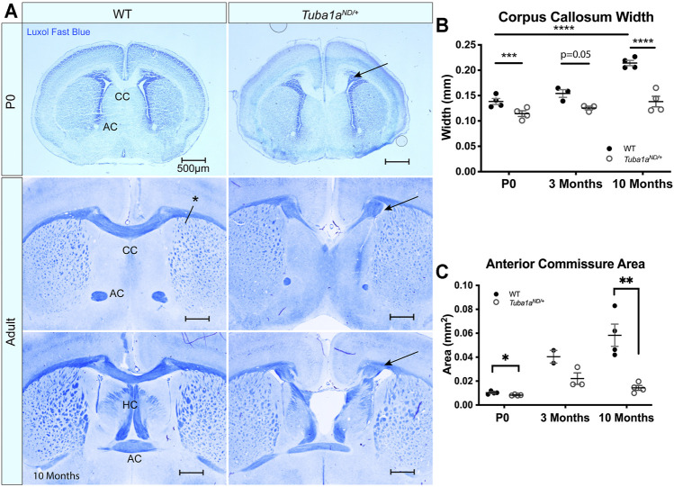
FIGURE 3.
Tuba1a is required for formation of midline commissural structures. (A) Luxol fast blue-stained coronal brain sections from postnatal day 0 (P0; top) and Adult (middle-bottom) wild-type and Tuba1a ND/+ mice. Images portray abnormal midline commissural formation in Tuba1a ND/+ mouse brains, with labels highlighting the corpus callosum (CC), anterior commissures (AC), and hippocampal commissure (HC). Scale bars are 500 μm. Arrows indicate Probst bundles. Asterisk in A. shows where measurements for B. were obtained. (B) Scatter plot representing corpus callosum width at P0, 3 months, and 10 months-old. (C) Scatter plot displaying anterior commissure area in P0, 3 months, and 10 months-old wild-type and Tuba1a ND/+ brains. Wild-type and Tuba1a ND/+ measurements compared by two-way ANOVA. *p < 0.05; **p < 0.01; ***p < 0.001; and ****p < 0.0001.

