Fig. 5.
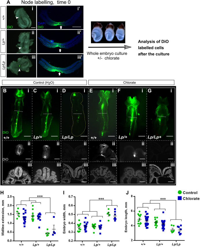
Chlorate prevents Closure 1 without affecting midline extension of Lp/+ embryos. (A) The node of E8.5 embryos from Vangl2Lp/+×Vangl2Lp/+ matings (0- to 5-somite stage, blind to genotype) was labelled by microinjection of the lipophilic fluorescent dye (DiO) (A-i,ii,iii). Embryos were then randomly allocated to 10 mM chlorate or water treatment and cultured for 24 h. Transverse sections through embryos at time 0 show the node and floor plate are successfully labelled with DiO in all three genotypes (A-i′,ii′,iii′). (B-G) Ventral view (rostral to the top) and transverse sections (trunk region) of DiO-injected embryos, after 24 h culture. Control (water) and chlorate-treated +/+ and Lp/+ embryos (B,C,E,F) exhibit marked midline extension of DiO-labelled cells, as detected in both whole mount and sections. Sections reveal failed Closure 1 in chlorate-treated Lp/+ embryos (F-ii,iii). In contrast, Lp/Lp embryos from both treatment groups display very limited midline extension of DiO-labelled cells (D-i,G-i), and fail in Closure 1 (D-ii,iii,G-ii,iii). Note that DiO labelling of the cranial region is non-specific, due to release of DiO into the amniotic cavity during labelling. (H) Midline extension measurements: points represent the distance DiO-labelled cells have extended along the caudal-to-rostral axis in individual embryos, with mean±s.e.m. also shown. Lp/Lp embryos exhibit significantly less midline extension than other genotypes. Chlorate-treated Lp/+ embryos do not differ from +/+ (water or chlorate) or Lp/+ (water) groups. (I,J) Embryo width (I) and length (J) measurements reveal a significantly wider and shorter body axis in Lp/Lp embryos than in other genotypes, irrespective of water/chlorate treatment. Width and length of Lp/+ embryos do not differ significantly between water and chlorate groups, nor do these values differ from +/+ embryo measurements. Data points are individual measurements, with mean±s.e.m. values also shown. *** P<0.001 (two-way ANOVA). Scale bars: 50 µm (B-ii,iii,C-ii,iii,D-ii,iii,E-ii,iii,F-ii,iii,G-ii,iii); 100 µm (A-i′,ii′,iii′); 200 µm (A-i,ii,iii); 0.5 mm (B-i,C-i,D-i,E-i,F-i,G-i).

