Fig. 6.
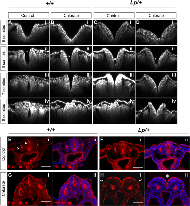
Chlorate alters neural plate morphology but not overall F-actin distribution in cultured embryos. (A-D) Embryos were cultured for 8 h with addition of 10 mM chlorate or water as control, fixed, stained with CellMask™ and imaged using confocal microscopy for morphological analysis. Images were re-sliced in Fiji to obtain transverse sections of the Closure 1 region, at the level of the 3rd somite. +/+ (A,B) and Lp/+ (C,D) embryos with 5, 6, 7 or 8 somites are shown (i-iv for each genotype/treatment combination). Closure 1 is normally completed from the 6-somite stage onwards. In control (water-treated) +/+ embryos, the neural plate adopts an increasingly horseshoe shape, concave inwards, with fusion evident dorsally from 7 somites (A). Chlorate delays this transition in +/+ embryos, with an initially V-shaped morphology, but closure is achieved by 8 somites, when the neural tube appears largely normal (B). Water-treated Lp/+ embryos resemble chlorate-treated +/+ embryos, and achieve closure by 8 somites (C). Chlorate-treated Lp/+ embryos exhibit a persistently V-shaped neural plate with convex curvature, in which the dorsal aspects of the neural folds fail to converge and fusion fails (D). Asterisks in A-ii, B-ii and C-ii indicate sites of initial contact between neural folds. (E-H) Phalloidin staining to detect F-actin distribution in transverse sections of the Closure 1 region of embryos cultured for 24 h. F-actin is enriched at the apical surface of NE (arrow in E-i) and apically within the epithelial somites (arrowhead in E-i). Although failure of NT closure is seen in chlorate-treated Lp/+ embryos (asterisk in H-ii), the only obvious difference from +/+ (E,G) and water-treated Lp/+ embryos (F) is an apparently reduced intensity of phalloidin staining at the lateral edges of the open neural folds (arrows in H-i) (n=4 embryos each). Scale bars: 50 µm (A-D); 100 µm (E-H).

