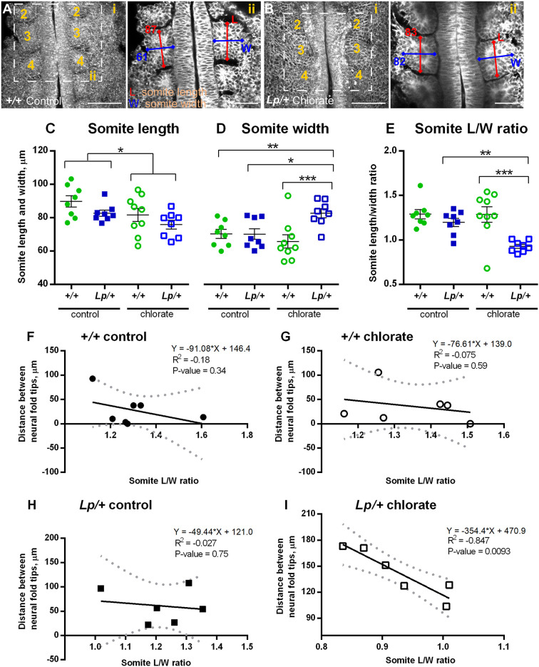
Fig. 7.
Altered somite morphology and correlation with Closure 1 delay in Lp/+ embryos after chlorate treatment. Embryos were cultured for 8 h from 0- to 5-somite stage, with addition of 10 mM chlorate or water as control, and prepared for morphological analysis as in Fig. 6A-D. (A-i,B-i) Z-projections of water-treated +/+ embryo (A-i) and chlorate-treated Lp/+ embryo (B-i), both at the 7-somite stage. Rostral is to the top; somites numbered sequentially. Images were re-sliced in Fiji (from dorsal to ventral surface) to obtain horizontal sections through the somite row. (A-ii,B-ii) Single z-planes through the Closure 1 region of the same embryos as in A-i and B-i. Length (L; rostrocaudal dimension) and width (W; mediolateral dimension) measurements were taken half way through the somite, at the level of the 3rd and 4th somites. Two somite pairs (4 individual somites) were measured per embryo. The L and W measurements (in µm) of the 3rd somite are shown for both embryos. (C-E) Somite length, width and L/W ratio in control and chlorate-treated +/+ and Lp/+ embryos, at the 6- to 7-somite stage. Individual points on the graphs are measurements averaged over 4 somites for each embryo, with mean±s.e.m. of embryo replicates. Note that somite width is significantly increased, and L/W ratio significantly reduced, in chlorate-treated Lp/+ embryos. *P<0.05; **P<0.01; ***P<0.001 (two-way ANOVA). (F-I) Linear regression analysis of distance between neural folds (µm) at the Closure 1 site and somite L/W ratios, with 95% confidence intervals (dotted lines) and a best fit line (solid lines). No significant correlation is detected for +/+ embryos, in either control (F) or chlorate (G) groups, nor for water-treated Lp/+ embryos (H). In contrast, chlorate-treated Lp/+ embryos (I) show a strong negative correlation (P=0.0093), with the greatest closure delay in embryos with the lowest somite L/W ratios (6- to 7-somite stage; n=6 in each case).

