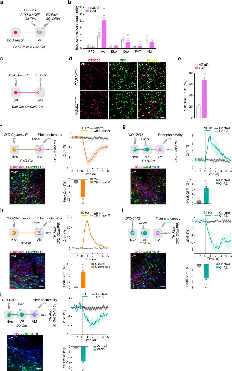
Fig. 5. D1NAc-VM and D1NAc-VP pathways drive opposing regulation of VM DAergic and GABAergic neurons.
a, b Schematic of rabies virus-based monosynaptic tracing (a) and quantification of input connection strength index (b). RV-ENVA-deltaG-dsRed (RVdG) was injected into the VP 2-week after unilateral injection of AAV9-EF1α-DIO-his-eGFP-2a-TVA, and AAV9-EF1α-DIO-RVG into the VP of Gad-Cre and vGlut2-Cre mice. Numbers of labeled presynaptic neurons/numbers of starter neurons were plotted [Two-tailed Student’s t-test, Gad n = 4, vGlut2 n = 4, NAc t(6) = 3.318, P = 0.016] *P < 0.05. c–e CTB retrograde labeling of GABAVP-VM and vGlut2VP-VM neurons. CTB555 was injected into the VM and AAV9-EF1α-DIO-H2B-GFP was injected into the VP of Gad-Cre or vGlut2-Cre mice (c). Representative images of retrograde labeling (d). Percentage of GABAVP-VM and vGlut2VP-VM neurons (e) [Two-tailed Student’s t-test, Gad n = 5, vGlut2 n = 3, t(6) = −17.902, P = 0.000002.] ***P < 0.001. f–j Optical fibers were implanted in the VM/VP and VM to exert laser stimulation and record CaMP6s signal simultaneously. Confocal images show ChrimsonR+ or ChR2+ terminals (Red), GCaMP6s+ cell bodies (Green), and TH+ neurons (Blue) in the VM. Scale bar: 50 μm. Statistical graph of group average GCaMP responses aligned to the onset of optical stimulation and bar graph of peak response to optical stimulation at 20 Hz. AAV9-hSyn-FLEX-ChrimsonR-tdTomato or AAV9-EF1α-DIO-hChR2-mCherry was injected into the NAc and AAV9-EF1α-DIO-GCaMP6s was infected in the VM of Gad-Cre mice. Calcium activity of VM GABAergic neurons in response to optical stimulation of GABANAc-VM or GABANAc-VP projection was recorded (f, g). [f Control n = 5, ChrimsonR n = 5, Two-tailed Student’s t-test, t(8) = 7.014, P = 0.000111; g Control n = 5, ChR2 n = 5, Mann-Whitney U test, Z = 2.611, P = 0.0079] **P < 0.01 and ***P < 0.001. AAV9-hSyn-FLEX-ChrimsonR-tdTomato or AAV9-EF1α-DIO-hChR2-mCherry was infected into the NAc and AAV9-TH-FlpO and AAV9-EF1α-fDIO-GCaMP6s were injected into the VM of D1-Cre mice. Activity of VM DAergic neurons in response to optical stimulation of D1NAc-VM or D1NAc-VP projection was recorded (h, i). [Mann-Whitney U test: h Control n = 6, ChrimsonR n = 6, Z = 2.882, P = 0.0022; i Control n = 6, ChR2 n = 6, Z = −2.882, P = 0.0022] **P < 0.01. (j) AAV9-EF1α-DIO-hChR2-mCherry was injected into the NAc and AAV9-TH-FlpO and AAV9-EF1α-fDIO-GCaMP6s were injected into the VM of D2-Cre mice. Activity of VM DAergic neurons in response to optical stimulation of D2NAc-VP projection was recorded. [Mann-Whitney U test, Control n = 6, ChR2 n = 6, Z = −2.882, P = 0.0022] **P < 0.01.

