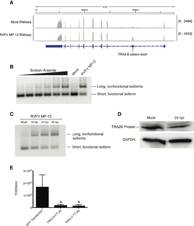
Figure 5.

TRA2-β mRNA is alternatively spliced in response to RVFV infection. (A) IGV tracks of mock- vs. RVFV MP-12-infected HEK293 cells at MOI = 1, highlighting TRA2-β. TRA2-β poison exon is labeled. (B) HEK293 cells were treated with sodium arsenite in increasing amounts (0, 1, 10, 25, 50, or 100 μM) or infected with RVFV MP-12 for 24h. RT-PCR with primers targeting TRA2-β mRNA was visualized by agarose gel. (C) HEK293 cells were infected with RVFV MP-12 at MOI = 1 for the times indicated, and RT-PCR against TRA2-β was performed. (D) HEK293 cells were infected with RVFV for 24h at MOI = 1, then cells were lysed and visualized via western blot. (E) TCID50 results obtained by infecting naïve Vero cells using RVFV MP-12 particles obtained from HEK293 cells transfected with indicated plasmids and subsequently infected with RVFV MP-12 at MOI = 1. Asterisks indicate p < 0.05 compared to GFP transfection (one-way ANOVA followed by Tukey’s HSD).
