Fig. 2.
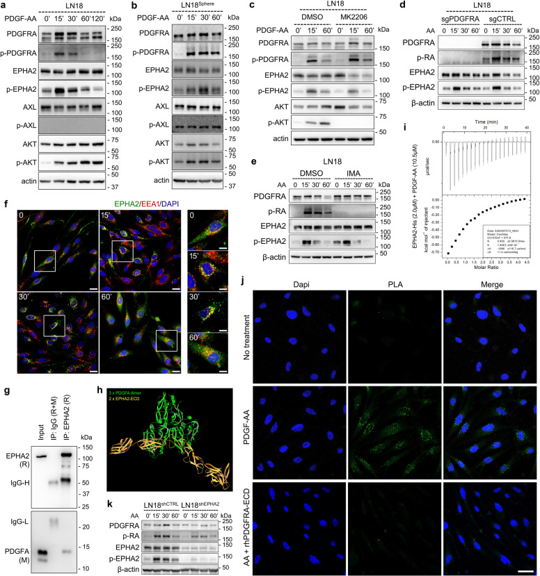
PDGFA activates EPHA2 in a PDGFRA-independent manner in GBM cells. a PDGFA-induced temporal expression of indicated proteins in LN18 cells examined by western blotting. β-actin is used as loading control. b PDGFA-induced temporal expression of indicated proteins in LN18 Sphere examined by western blotting. c PDGF-A-induced temporal expression of indicated proteins in LN18 cells pre-treated with DMSO and MK2206. d PDGFA-induced temporal expression of indicated proteins in LN18 cells infected with lentivirus containing control sgRNA or sgRNA targeting PDGFRA. e PDGFA-induced temporal expression of indicated proteins in LN18 cells pre-treated with vehicle or IMA. f Representative immunofluorescence images stained by antibodies targeting EPHA2 and EEA1, respectively. DAPI is used to label nuclei. Scale bar = 10 μm for large four panels and 5 μm for small four panels. g Co-immunoprecipitation and western blotting of EPHA2 in PDGFA-treated LN18 cells. h Interaction simulation of three-dimension structure of PDGFA and EPHA2 extracellular domain. i Interaction thermodynamics of recombinant human EPHA2 extracellular domain and recombinant human PDGF-AA using Microcal iTC200. j Proximity ligation assay using LN18 cells without treatment, treated with PDGFA for 15 min, or pre-treated with recombinant PDGFRA extracellular domain followed by PDGFA treatment for 15 min. The cells is counterstained with Dapi (blue) to mark nuclei. Green dot signals represent interaction between EPHA2 and PDGFA. Scale Bar = 25 μm. k PDGFA-induced temporal expression of indicated proteins in LN18 cells infected with lentivirus containing control shRNA or shRNA targeting EPHA2.

