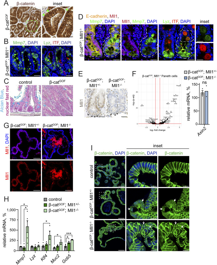
Figure S3. Mll1-deficient β-catGOF crypts and organoids exhibit goblet cell features.
(A) Representative immunohistochemistry staining for β-catenin on section of β-catGOF intestine, nuclei counterstained with haematoxylin, scale bar 50 μm. Magnification of inset on the right, scale bar 25 μm. Arrowheads (green) mark cells with nuclear β-catenin. Stainings were performed in n = 3 independent mice. (B) Representative immunostainings for the Paneth cell markers Mmp7 (green, left) and Lyz (green, right) and the goblet cell marker ITF (red) on serial sections of β-catGOF; Mll1+/− intestine, nuclei in blue (DAPI), scale bars 50 μm. Stainings were performed in n = 3 independent mice. (C) Alcian blue stainings for goblet cells on sections of uninduced control and β-catGOF intestine. Counterstaining with nuclear fast red, scale bars 50 μm. (D) Immunostainings on sections of adjacent non-recombined (left) and recombined mutant (right) small intestinal crypts in β-catGOF; Mll1−/− mice, left: for Mmp7 (green) and Mll1 (red), right: for Lyz (green) and ITF (red), scale bars 25 μm. Insets on the right: magnifications of mutant double-positive cells, scale bars 10 μm. Nuclei stained with DAPI (blue), E-cadherin (yellow) stains cell borders. Stainings were performed in n = 3 independent mice. (E) Immunohistochemistry for Mll1 on sections of small intestinal crypts in β-catGOF; Mll1+/− and β-catGOF; Mll1−/− mice at 10 d after the induction of mutagenesis, nuclei counterstained with haematoxylin, scale bars 25 μm. Crypts surrounded by dashed lines, nuclei of vesicle-containing Paneth cells at the crypt base marked by arrowheads. (F) Left: volcano plot of differentially expressed genes in Paneth cells isolated from β-catGOF; Mll1+/− and β-catGOF; Mll1−/− intestines at 10 d after induction of mutagenesis, n = 4 independent mice per genotype. Cutoffs log2 fold-change ≥0.5, adjusted P-value (P-adj) ≤ 0.05. Right: mRNA expression of Axin2 in Paneth cells isolated from the intestines of n = 3 β-catGOF; Mll1+/− and n = 2 β-catGOF; Mll1−/− independent mice at 10 d post induction. Data are presented as mean values ± SEM. (G) Immunostaining for Mll1 (red) on sections of β-catGOF; Mll1+/− and β-catGOF; Mll1−/− organoids, nuclei in blue (DAPI), scale bars 50 μm. (H) mRNA expression of secretory cell genes Mmp7, Lyz, Klf4, Muc2, and Gob5 in non-induced control, β-catGOF; Mll1+/− and β-catGOF; Mll1−/− organoids relative to β-catGOF organoids, n = 3 independent experiments, two-tailed unpaired t test, Mmp7: *P = 0.049 (control - β-catGOF; Mll1−/), *P = 0.04 (β-catGOF; Mll1+/− - β-catGOF; Mll1−/−), Klf4: *P = 0.04, Muc2: *P = 0.02, Gob5: *P = 0.04, ***P = 0.0006. Data are presented as mean values ± SEM. (I) Immunostainings for β-catenin (green) on sections of non-induced control, β-catGOF; Mll1+/− and β-catGOF; Mll1−/− organoids, scale bars 50 μm. Magnifications in insets, scale bars 10 μm. DAPI (blue) stains nuclei.

