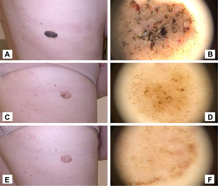
Figure 7.
Case 1. (A and B) Macro-photo and dermoscope pictures of a large and thick seborrheic keratosis before HIFU. (C and D) Macro-photo and dermoscope pictures 6 weeks after initial HIFU treatment. The seborrheic keratosis has clearly reduced in thickness and the majority of brown clods and coiled vessels have been removed. The lesion is however still clearly visible, in particular in the periphery of the original lesion. A second repeat treatment was administered. (E and F) Macro-photo and dermoscope picture at follow-up visit 10 weeks after first treatment (4 weeks after second treatment). The visual appearance is significantly improved, but seborrheic keratosis is still visible, and a third treatment will be needed to fully remove the lesion.

