Figure 1.

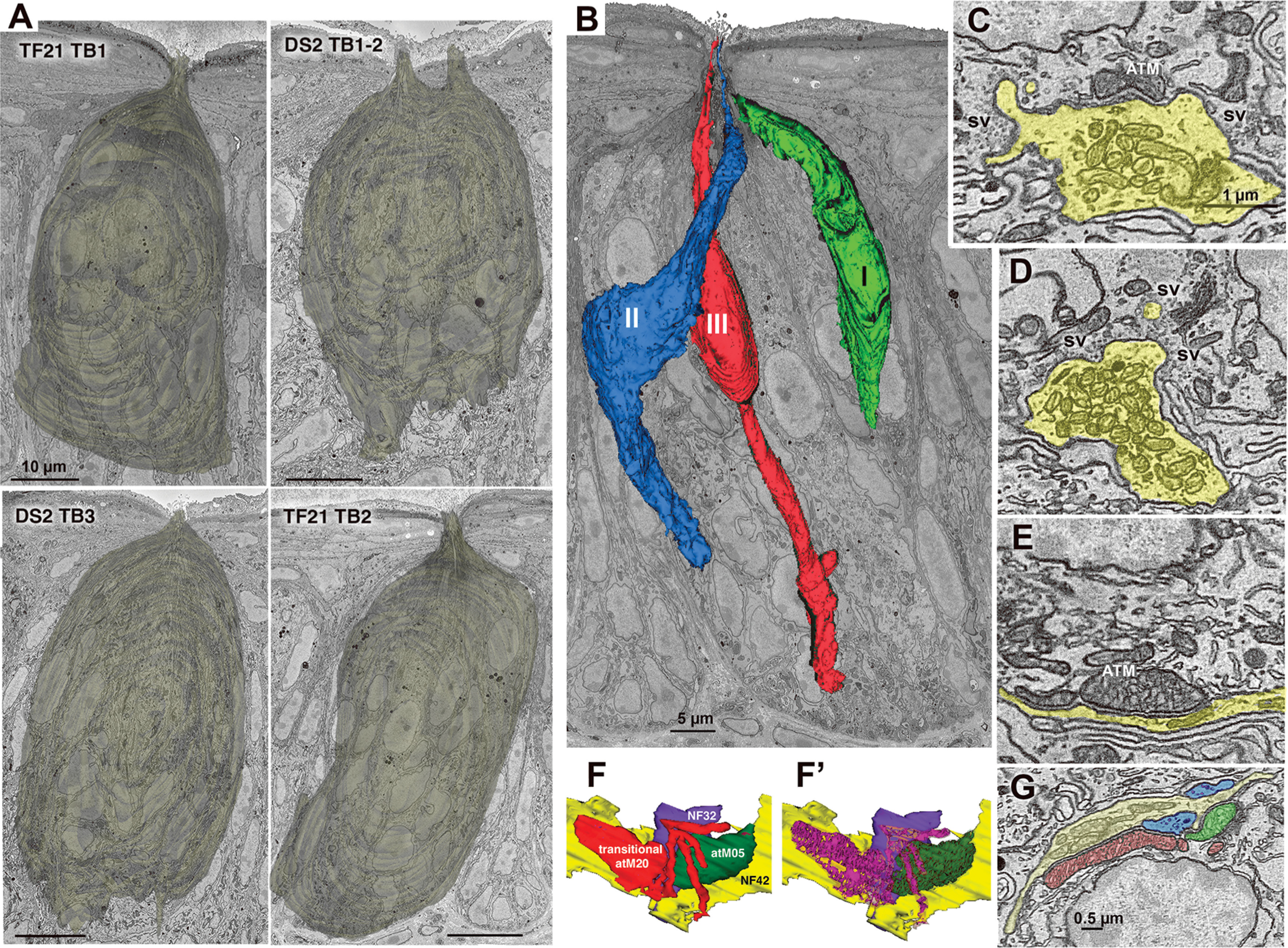
Taste buds, cell types, and synapses. A, Reconstructions of the appearance of the five segmented taste buds in the connectome dataset. The upper right image (DS2_TB1-2) shows the conjoined taste buds with two distinct apical pores. Scale bars: 10 µm. B, Taste bud TF21_TB2 showing representatives of the three elongate cell types. C, Hybrid type synapse showing both an ATM and a cluster of synaptic vesicles (sv) at the region of contact between the NF (yellow) and Type III taste cell. D, Vesicular synapse characteristic of Type III taste cells. Numerous, densely packed conventional mitochondria are contained within the nerve process (yellow). E, Channel synapse characteristic of Type II cells with ATM apposed to the plasma membrane at the region of contact with the NF. F, F', 3D reconstructions of a transitional mitochondrion at a synapse between a Type II cell and two NFs (blue and yellow). The transitional mitochondrion (red) with mixed lamellar and tubular cristae (magenta, F') is adjacent to an ATM (green, F) containing only tubular cristae (dark green, F'). G, EM image of the mitochondrial complex reconstructed in panel F.
