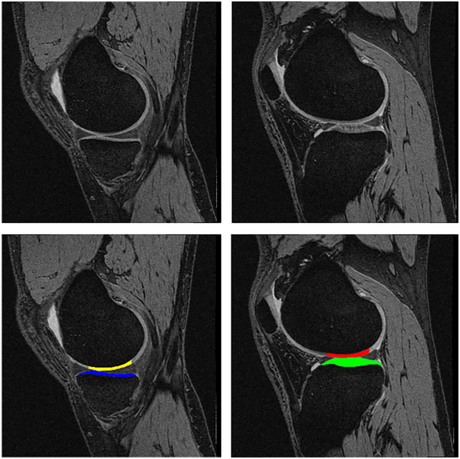
Figure 1.
Sagittal DESS MRI sequence displaying the medial femorotibial compartment (MFTC) on the left and the lateral one (LFTC) on the right. Top row without segmentation, bottom row with segmentation of the cartilages. Yellow = weight-bearing medial femorotibial condyle (cMF); blue = medial tibia (MT); red = weight-bearing lateral femorotibial condyle (cLF); green = lateral tibia (LT).

