
Figure 2.
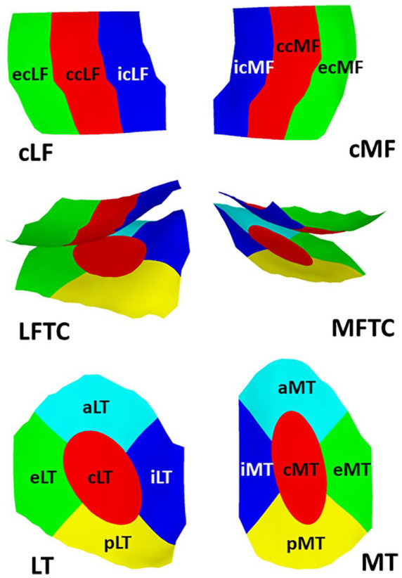
Explosion figure displaying the 16 femorotibial subregions. Top row: Inferior view of the weight-bearing femorotibial condyles, with the lateral weight-bearing (central) femorotibial condyle on the left, and the medial one on the right. Bottom row: Superior view of the tibial plateau, with the lateral tibia on the left, and the medial one on the right. The middle row shows a posterior view of the femoral condyles and tibiae together. Red = central subregions (c); green = external subregions (e); blue = internal subregions (i); yellow = posterior subregions (p); turquoise = anterior subregions (a).