Figure 3.
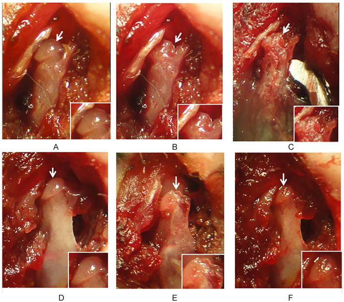
Evaluation of condylar cartilage damage (CCD) after mesenchymal stromal cell (MSC) transplantation. Condylar head of untreated CCD (A-C). The condylar cartilage is observed before (A, arrow) and after CCD (B, arrow). Chondral defect after 6 weeks (C). The chondral defect remains without evidence of tissue repair (arrow and insert). Condylar head of MSC-treated CCD (D-F). The condylar cartilage is observed before CCD (D, arrow). Chondral defect after CCD (E, arrow), and after 6 weeks post-MSC transplantation (F, arrow). Evidence of cartilage regeneration is observed at the site of CCD were MSC were implanted (F). New cartilage-like tissue is observed filling the chondral defect (arrow and insert). Results are representative of 4 CCD (one joint in each TMJ ) in each group, all of which had similar results.
TMJ = temporo mandibular joint.

