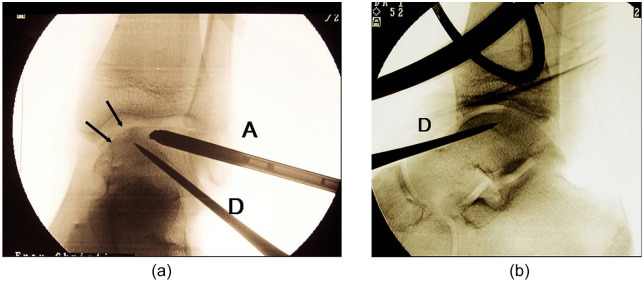
Figure 1.
(a) Intraoperative fluoroscopy during arthroscopy of the ankle joint and simultaneous retrograde drilling of a talar osteochondritic lesions (anteroposterior view). Arrow = talar lesion. (b) Intraoperative fluoroscopy during arthroscopy of the ankle joint and simultaneous retrograde drilling of a talar osteochondritic lesions (anteroposterior view). A = arthroscope; D = drill.

