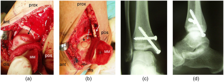
Figure 2.
(a) Intraoperative findings of a medial osteochondritis dissecans (OCD) lesion of the talus after medial malleolar osteotomy (arrow = lesion, ant = anterior, prox = proximal, pos = posterior, dis = distal, MM = medial malleolus after osteotomy). (b) Intraoperative findings of a medial OCD lesion of the talus after medial malleolar osteotomy (arrowhead = osteochondral autograft transplant in the anterior part of the lesion, gray arrow: remaining posterior part of the lesion to be transplanted; ant = anterior, prox = proximal, pos = posterior, dis = distal, MM = medial, malleolus after osteotomy, white arrow: retromalleolar tendons). (c) Postoperative x-ray (anteroposterior view) showing the ankle joint after refixation of the medial malleolus with 2 screws. (d) Postoperative x-ray (lateral view) showing the ankle joint after refixation of the medial malleolus with 2 screws.

