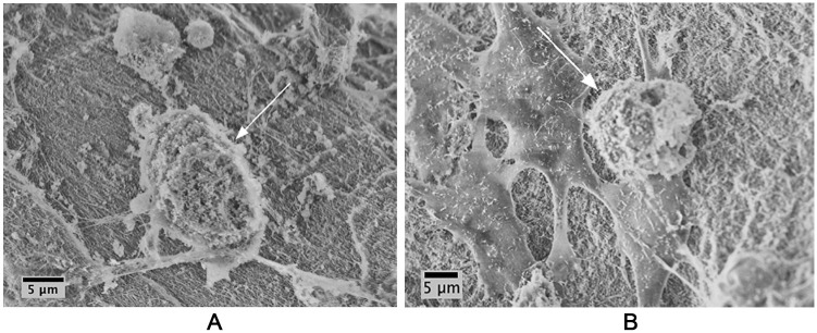
Figure 3.
Mesenchymal stem cells (MSCs) and chondrocytes adhere and migrate on BioCartilage. (A) MSCs adhere to and spread (arrow) on BioCartilage; (B) chondrocytes adhere to and migrate (arrow) on BioCartilage. BioCartilage was mixed with MSCs or chondrocytes in platelet-poor plasma (PPP) and grafted into cartilage defects to model cartilage repair. Repair constructs were cultured for 48 hours, then fixed and dehydrated prior to scanning electron microscopy (SEM) imaging. SEM was conducted at an accelerating voltage of 5 keV and an aperture size of 30 μm.

